Bronchi vs Bronchioles: What’s the Difference?

It’s no secret that mammals breathe through their lungs. Their respiratory system comprises the bronchioles, mouth, larynx (voice box), bronchi, alveoli, and trachea. The bronchioles and bronchi are tubular structures. While the bronchi constitute C-shaped cartilages, the bronchioles lack cartilaginous support.
Furthermore, bronchi have a larger diameter compared to bronchioles as they’re located before the respiratory passageway. Bronchi stem from the trachea, forming bronchioles that are linked to the alveoli. The major difference between bronchioles and bronchi is that the latter is involved in the cleaning, conducting, and warming of the air in the respiratory passageway.
Bronchioles are involved in gas exchange in addition to the conduction of air. In this guide, we’ll explore other differences between bronchi vs bronchioles.
What Are Bronchi?
Bronchi can be defined as the tubules that form the main passageway of air into the lungs. The air makes its way from the nose to the trachea and larynx. The trachea conducts air to bronchi that decrease in diameter when they come from primary to tertiary. Below are the three categories of bronchi, categorized by how they branch.
Primary
These bronchi are found at the point of separation of the trachea into the right and left bronchus.
Secondary
These bronchi are found right at the center of your lungs.
Tertiary
Found near the bottom of your lungs, tertiary bronchi are right above the respiratory bronchiole.
The main role of the bronchi is the conduction of air to the bottom of your lungs. The wall of the bronchi comprises a fibrocartilage layer that curbs the bronchi tube from shrinking with every breath you take. It’s also lined with ciliated pseudostratified epithelium that is made up of mucus-secreting goblet cells.
The mucus is undefeated in trapping pathogens and dust, whereas the cilia eliminate the dust from the respiratory passageway. Nonetheless, no exchange of gases occurs through the wall of the bronchi. During conduction, the air is cleaned and warmed.
As a result of infections, the swollen bronchi trigger bronchitis, which can be caused by excess mucus and phlegm, making breathing an uphill battle.
What Are Bronchioles?
These refer to the branches that form alveoli. Each lung has about 30,000 bronchioles and is not encircled with cartilages. Bronchioles comprise simple cuboidal epithelium with goblet cells, smooth muscle cells, and elastic connective tissue.
Based on the branching pattern, bronchioles fall into three categories.
Lobular Bronchioles
What differentiates these from the larger tubes is that the absence of cartilage and cubical-shaped ciliated epithelial cells. Also known as pre-terminal bronchioles, lobular bronchioles are about 0.2mm in diameter.
Each bronchiole branches into at least two respiratory bronchioles (terminal bronchioles) with scattered alveoli after passing into a pulmonary lobule. They further branch into various alveolar ducts with more alveoli connected to them.
Terminal Bronchioles
These represent the most distal part of the conducting zone. Terminal bronchioles branch off the lesser ones, with each bronchiole dividing to form respiratory bronchioles containing few alveoli. Lined with simple cuboidal epithelium, terminal bronchioles contain club cells.
Furthermore, they have a limited number of ciliated cells and lack goblet cells. Club cells are protein-secreting, non-ciliated, and rounded. Their secretions, known as surfactant, are a proteinaceous, non-sticky compound that effectively maintains the airway in the tiniest bronchioles.
As a result, they diminish surface tension, paving the way for the bronchiole expansion during inhalation, preventing them from collapsing during exhalation. As stem cells of the respiratory system, club cells generate enzymes that detoxify dissolved substances in the respiratory fluid.
Respiratory Bronchioles
As the narrowest airways of the lungs, respiratory bronchioles measure 0.5mm in diameter. The bronchi divide a multitude of times before they evolve into the bronchioles, after which they supply air to the gas exchange surfaces of your lungs.
The alveoli interrupt the respiratory bronchioles. As continuations of the respiratory bronchioles, the alveolar ducts are lined by non-ciliated cells (Clara cells) coupled with ciliated columnar epithelium.
The two main functions of the bronchioles are:
- Gas Exchange
- Air Conduction To The Alveoli
The possible ailments in the bronchioles are:
Bronchitis
It defines the inflammation or swelling of the air passages that connect the lungs, nose, and mouth (bronchial tubes). Bronchitis causes symptoms such as difficulty in breathing, coughing, and wheezing.
Furthermore, you may have trouble clearing phlegm or heavy mucus from your airway. Bronchitis can be chronic or acute. While the latter usually clears up after a while, chronic bronchitis is persistent and never goes away entirely.
Bronchiectasis
It’s a lung condition that triggers coughing up mucus. In the lungs, the bronchi are the passages that permit air to enter the lungs. In Bronchiectasis, the insides of the bronchi become thicken after a while from inflammation that leaves scars. Mucus easily collects in these passages because the thicker walls are not sturdy enough to expel the mucus from the lungs. Additionally, the thin hair-like strands (cilia) are destroyed. Bronchiectasis can be characterized by coughing up blood or a lot of mucus, tightness or chest pain caused by the difficulty in breathing, clubbing of nails, and wheezing.
Bronchospasm
It’s the tightening of the muscles that line the bronchi (airways) in your lungs. Consequentially, your airways narrow which means they don’t allow as much air to go out of or come into your lungs. They limit the amount of oxygen entering your bloodstream and carbon dioxide being expelled from your blood. People with allergies or asthma are more susceptible to bronchospasm, characterized by shortness of breath and wheezing.
Bronchi Vs Bronchioles: Similarities
We’ve rounded up a few commonalities between the two.
- Bronchioles And Bronchi Are Two Types Of Tubular Structures That Are Found Before The Alveoli In The Respiratory Passageway
- The Epithelium Of The Bronchioles And Bronchi Are Made Up Of Goblet Cells Which Produce Mucus
- The Epithelium Of The Bronchioles And Bronchi Are Made Of Cilia
Bronchi Vs Bronchioles: What Are The Differences?
Let’s explore a few characteristics that differentiate the two.
1. Formation
Bronchioles: Formed from smooth muscle layers
Bronchi: Formed from the primary bronchi
2. Definition
Bronchioles: Refer to the branches that form alveoli
Bronchi: Refer to the tubules that create the main airway into the lungs
3. Form
Bronchi: Form bronchioles
Bronchioles: Form alveoli
4. Structure
Bronchioles: Constitute the epithelium, smooth muscles, and elastic fibers and tissues
Bronchi: Comprise a fibro-cartilaginous layer
5. Sections
Bronchioles: Can be broken down into respiratory, lobular, and terminal bronchioles
Bronchi: Can be categorized into tertiary, primary, and secondary bronchi
6. Cartilages
Bronchioles: Lack cartilaginous support
Bronchi: Comprise c-shaped cartilages that deliver excellent support
7. Type of Epithelium
Bronchioles: Constitute ciliated, simple cuboidal epithelium
Bronchi: Comprise the columnar, pseudostratified epithelium
8. Diameter
Bronchioles: Smaller in diameter compared to bronchi
Bronchi: Large diameter
9. Function
Bronchioles: The main functions are air conduction and gas exchanges
Bronchi: Their main responsibilities are air cleaning, warming, and conduction in the respiratory passageway
10. Air Conduction
Bronchioles: Conduct air to the alveoli
Bronchi: Conduct air to the bronchioles
11. Pathology
Bronchioles: Bronchospasm, bronchitis, and Bronchiectasis are the likely medical conditions
Bronchi: Bronchitis is the likely clinical condition
How To Take Care Of Your Lungs
Let’s discuss a few tips to keep your lungs in tiptop condition and live a healthier life.
Incorporate Greenery
Adding real plants to your home boosts the oxygen levels and quality of air. Their natural ability to get rid of certain toxins purifies the air, which is particularly useful when the harsh winters roll around, forcing you to spend most of your time indoors.
Adopt An Active Lifestyle
Exercise is ideal for your lungs as well. It strengthens your muscles and elevates blood circulation, two of the most essential requirements for the lungs to function perfectly. It’s for this reason exercise boosts their efficiency.
A Healthy Diet
It’s equally as excellent for your lungs as it is for other parts of your body. Studies have shown that low levels of important nutrients have been tied to various lung diseases. Consuming foods rich in magnesium, zinc, potassium, selenium, and vitamins E, A, and C help maintain a healthy respiratory system.
Water
Drinking plenty of water plays an important role in the health of your lungs. It aids in thinning the mucus secretions that naturally accumulate in the lungs every day, allowing you to breathe easier.
Steer Clear Of Tobacco Products
Chewing tobacco and smoking is harmful to your lungs and overall health. Breaking the habit of smoking and avoiding the use of tobacco products is one of the most effective ways to keep your respiratory system healthy.
Wash Your Hand
As obvious as it seems, washing your hands and practicing good hygiene is perfect for your lungs, more so during flu season and the ongoing pandemic. Doing so keeps bacteria, germs, and viruses that are detrimental to your lungs from entering your body.
To efficiently prevent those harmful microbes from making their way into your respiratory system, ensure you sanitize your telephone handsets, tablets, cell phones, computer keyboards, and other devices.
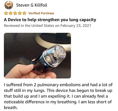
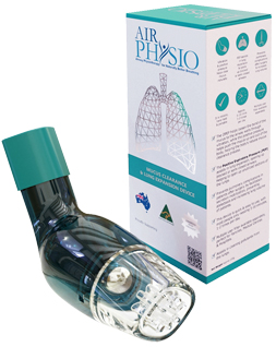
Average Lung AirPhysio
ENJOY BETTER BREATHING - Use this 100% Drug Free Device - AIRPHYSIO
Recent Posts
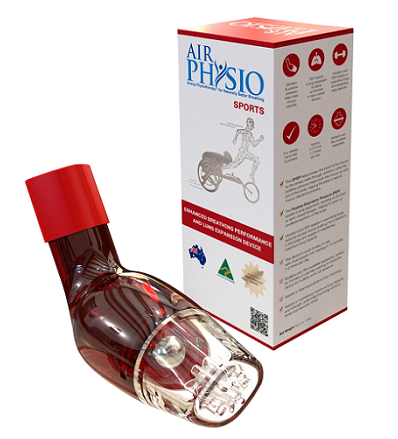
Sports AirPhysio
IMPROVE YOUR SPORTING PERFORMANCE - Use this 100% Drug Free Device - AIRPHYSIO
AirPhysio Child
BETTER BREATHING FOR YOUR CHILD - Use this 100% Drug Free Device - AIRPHYSIO
Categories
- asthma (2)
- atelectasis (2)
- bronchiectasis (2)
- copd (3)
- cystic-fibrosis (45)
- featured (10)
- uncategorized (2)